
Shiyun Liu, Mintian Zeng, Houlin Wang, Shenyu Miao & Jianhui Chen
Study on Female Gametophyte Development of Primulina tabacum
Botanical Research 2021, 10(1):35-46
ABSTRACT
The embryology of Primulina tabacum was studied by conventional paraffin section method to study the development regularity of female gametophyte, to understand the relationship between reproduction and the environment of the species, as well as the endangerment were also explored from the perspective of embryo development. Results showed the following: Anatropous ovule, parietal placentation, two carpels and one locule, tenuinucellate ovule, polygonum type embryosac. Various abortive phenomena occurred at different stages of megaspore development and female gametophyte formation. Its own reproductive ability was not strong, and various abnormal developments in the reproductive process, was one of the important reasons leading to its endangerment.
Original article link:
http://www.hanspub.org/journal/br;https://doi.org/10.12677/br.2021.101006
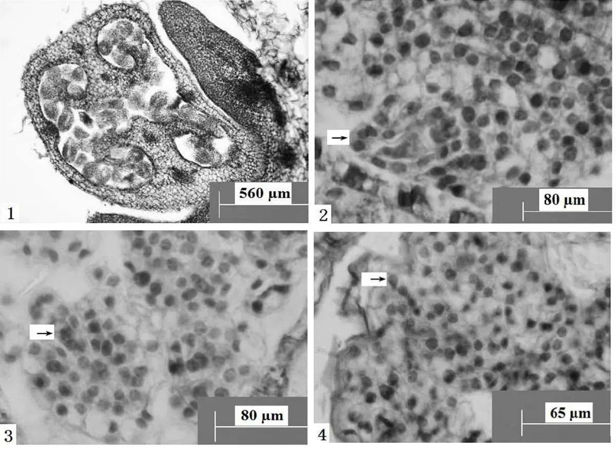
Figure 1. Transverse section of ovary, showing parieta placenta. Scale bars = 560 µm
Figure 2. Ovules longitudinal section, the arrow shows tenuinucellate ovule. Scale bars = 80 µm
Figure 3. Ovules longitudinal section, the arrow shows megaspore mother cells. Scale bars = 80 µm
Figure 4. Ovules longitudinal section, the arrow shows four megaspores arranged in a straight line. Scale bars = 65 µm
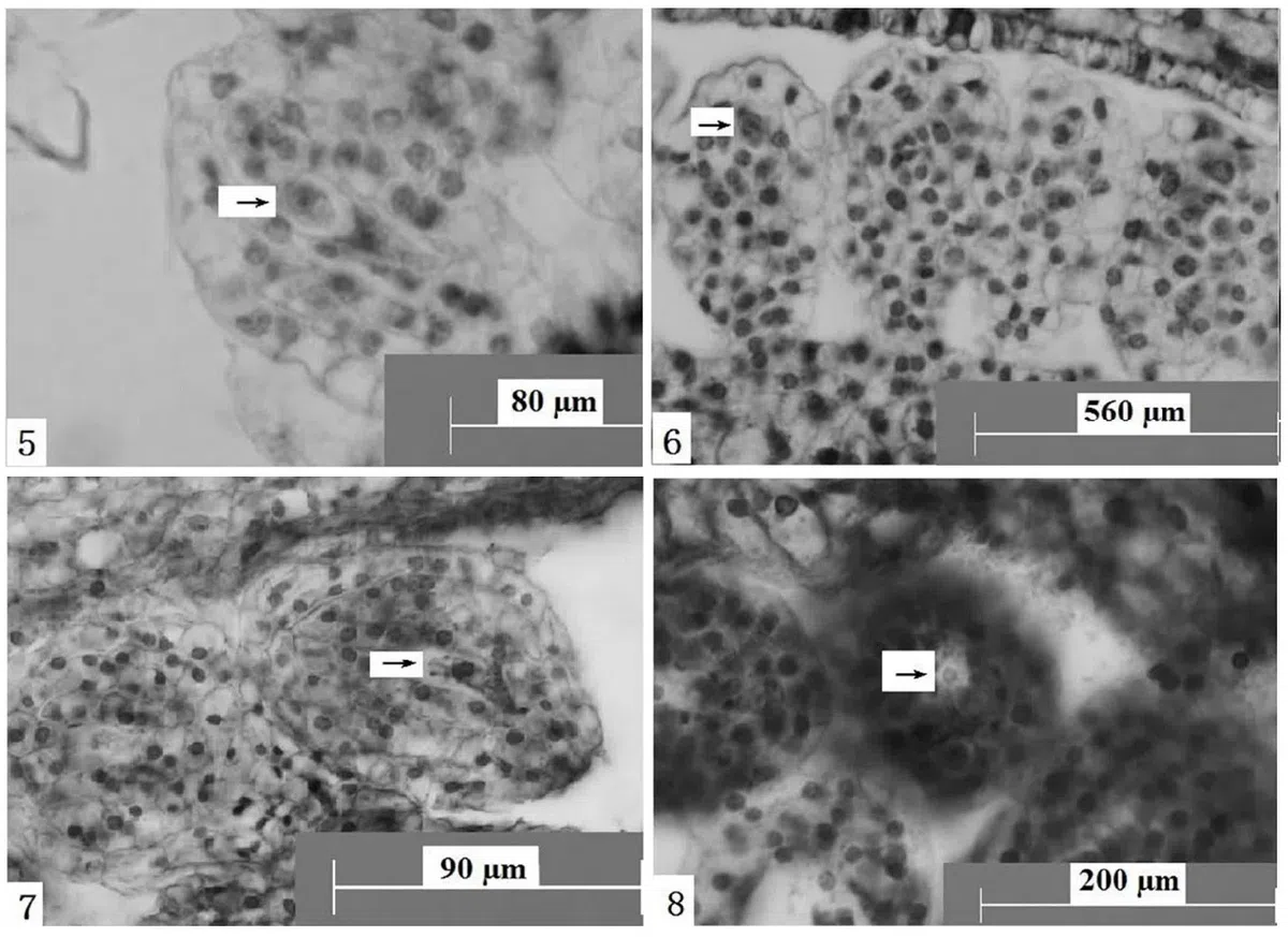
Figure 5. Ovules longitudinal section, the arrow shows functional megaspore. Scale bars = 80 µm
Figure 6. Ovules longitudinal section, the arrow shows megaspores arranged in a “T” shape. Scale bars = 560 µm
Figure 7. Ovules longitudinal section, the arrow shows disintegrate megaspore. Scale bars = 90 µm
Figure 8. Ovules longitudinal section, the arrow shows mononuclear embryo sac. Scale bars = 200 µm
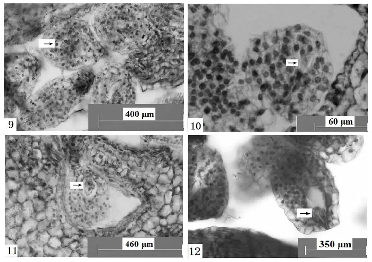
Figure 9. Ovules longitudinal section, the arrow shows two-nuclear embryo sac. Scale bars = 400 µm
Figure 10. Ovules longitudinal section, the arrow shows four-nuclear embryo sac. Scale bars = 60 µm
Figure 11. Ovules longitudinal section, the arrow shows eight-nuclear embryo sac. Scale bars = 460 µm
Figure 12. Ovules longitudinal section, the arrow shows egg apparatus. Scale bars = 350 µm
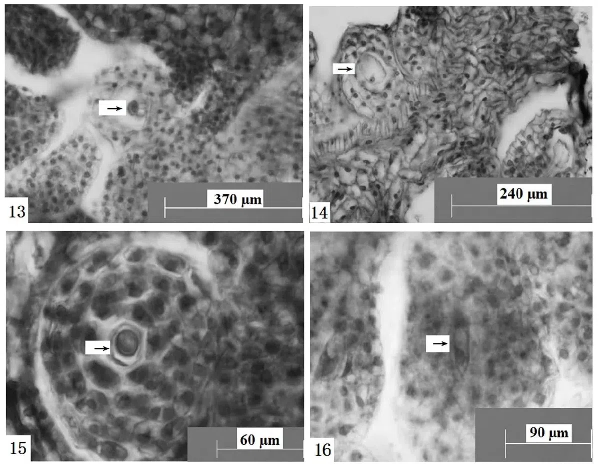
Figure 13. Ovules longitudinal section, the arrow shows antipodal cells. Scale bars = 370 µm
Figure 14. Ovules longitudinal section, the arrow shows central cell. Scale bars = 240 µm
Figure 15. Ovules longitudinal section, the arrow shows vacuolated mononuclear embryo sac. Scale bars = 60 µm
Figure 16. Ovules longitudinal section, the arrow shows disintegrated two-nuclear embryo sac. Scale bars = 90 µm
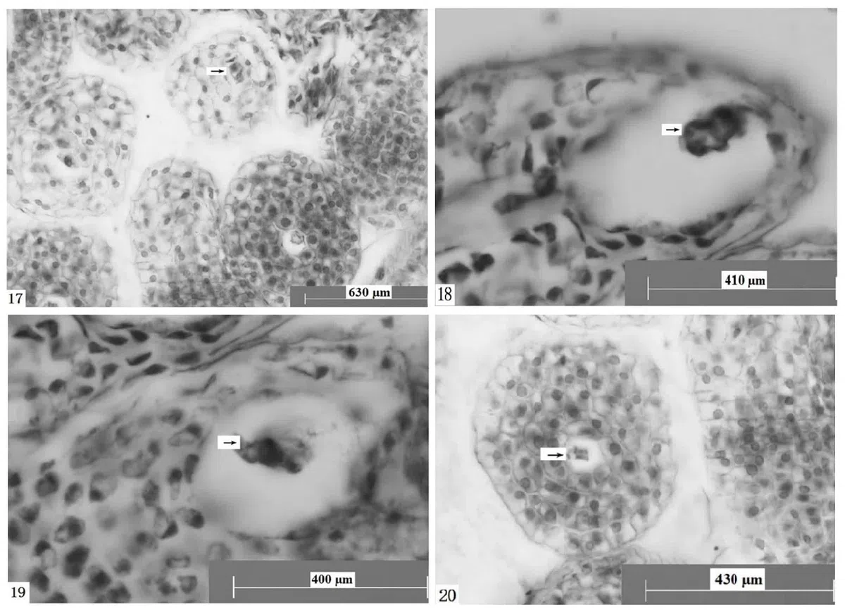
Figure 17. Ovules longitudinal section, the arrow shows disintegrated four-nuclear embryo sac. Scale bars = 630 µm
Figure 18. Ovules longitudinal section, the arrow shows polypolymeric cells clustered at the end of the pore. Scale bars = 410 µm
Figure 19. Ovules longitudinal section, the arrow shows a polymorphic body clustered in the center of the embryo sac. Scalebars = 400 µm
Figure 20. Ovules longitudinal section, the arrow shows a polymorphic body clustered in the center of the embryo sac. Scalebars = 430 µm
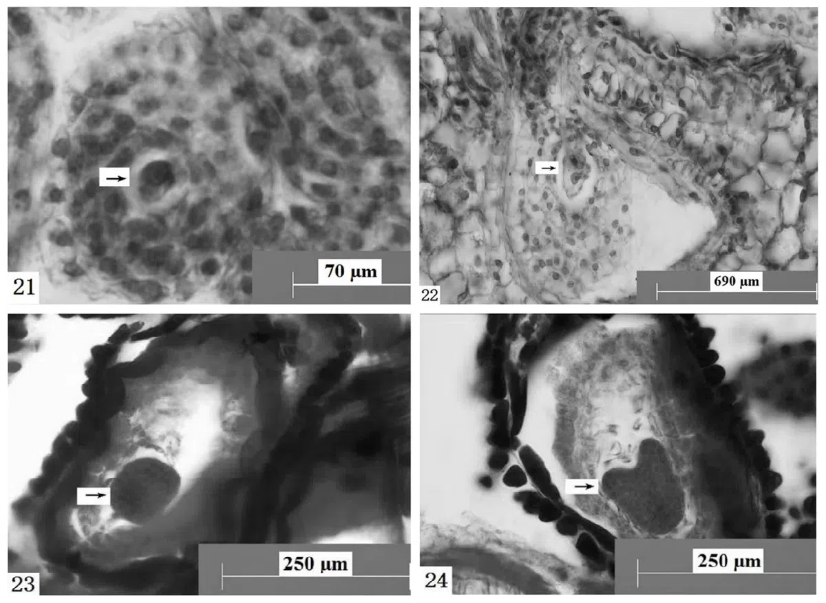
Figure 21. Ovules longitudinal section, the arrow shows sperm and egg apparatus cells. Scale bars = 70 µm
Figure 22. Ovules longitudinal section, the arrow shows sperm, egg cell, central cell. Scale bars = 690 µm
Figure 23. Ovules longitudinal section, the arrow shows globular embryo. Scale bars = 250 µm
Figure 24. Ovules longitudinal section, the arrow shows heart shaped embryo. Scale bars = 250 µm
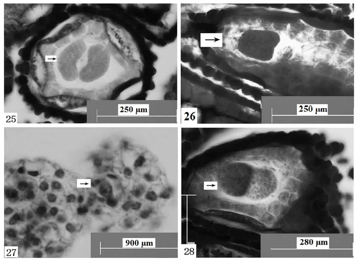
Figure 25. Ovules longitudinal section, the arrow shows cotyledon embryo. Scale bars = 250 µm
Figure 26. Ovules longitudinal section, the arrow shows a heart-shaped embryo with a suspensor. Scale bars = 250 µm
Figure 27. Ovules longitudinal section, the arrow shows endosperm nuclei and zygotes dividing. Scale bars = 900 µm
Figure 28. Ovules longitudinal section, the arrow shows embryo sac with double embryo. Scale bars = 280 µm
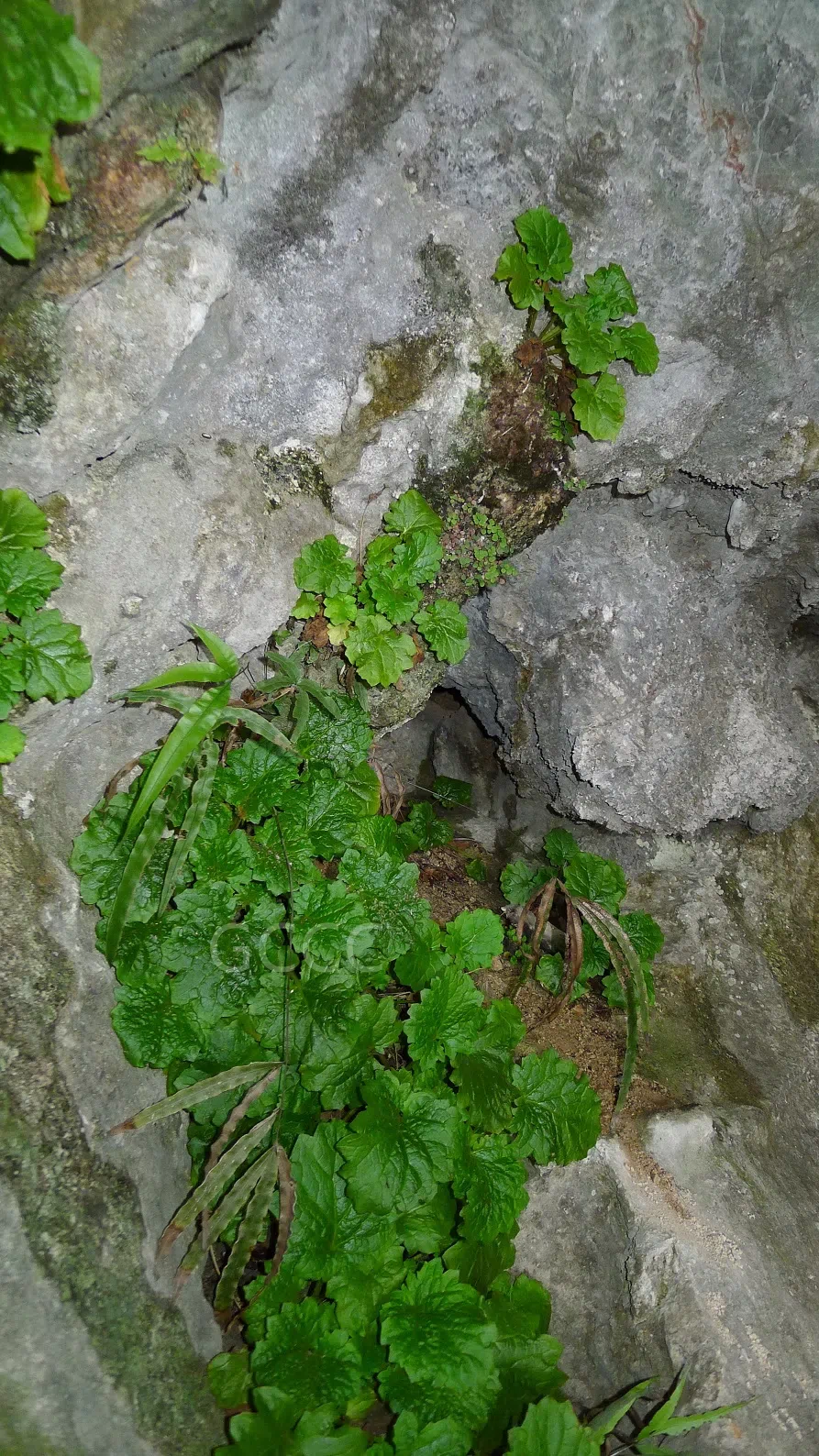
Figure 29.Primulina tabacum in the field
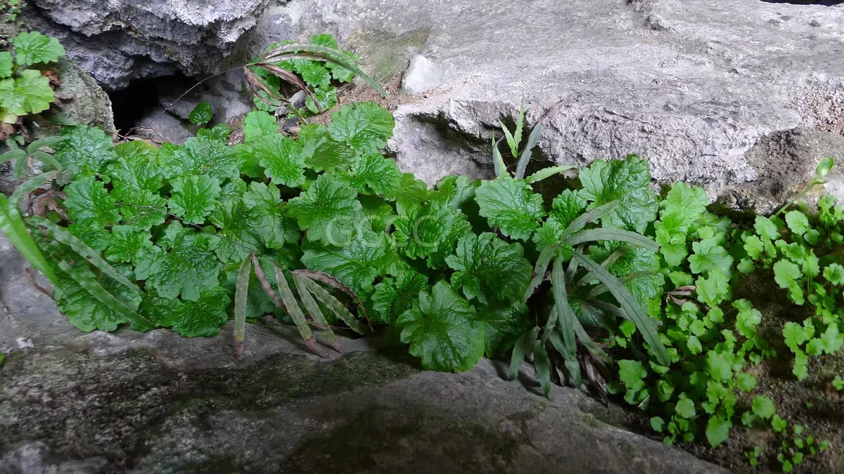
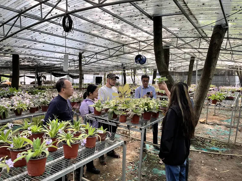
Figure 30. The growing status of Primulina tabacum and those seedlings in the field
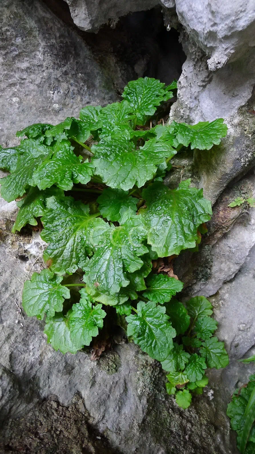
Figure 31. The mature individuals of Primulina tabacum in the field
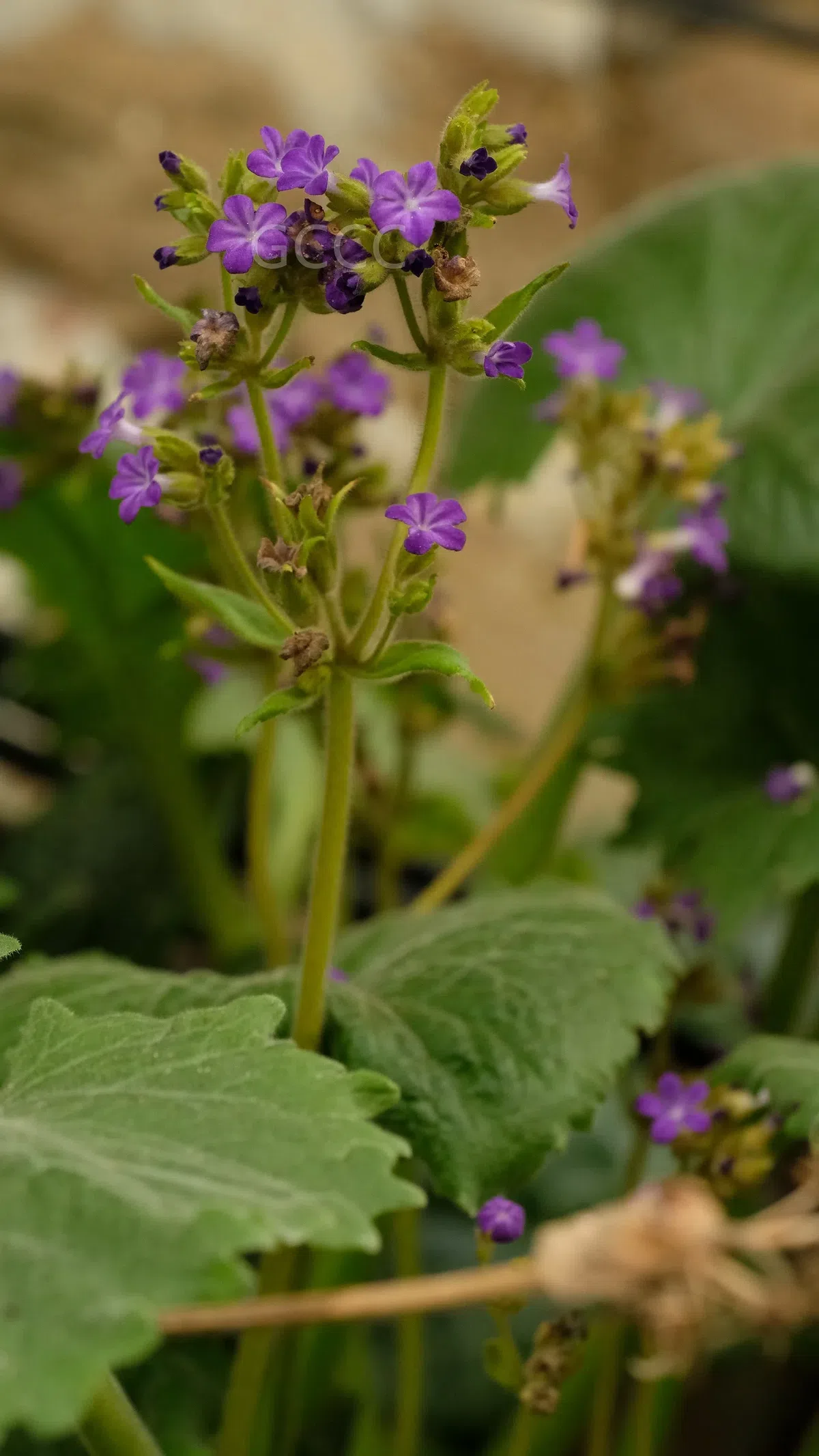
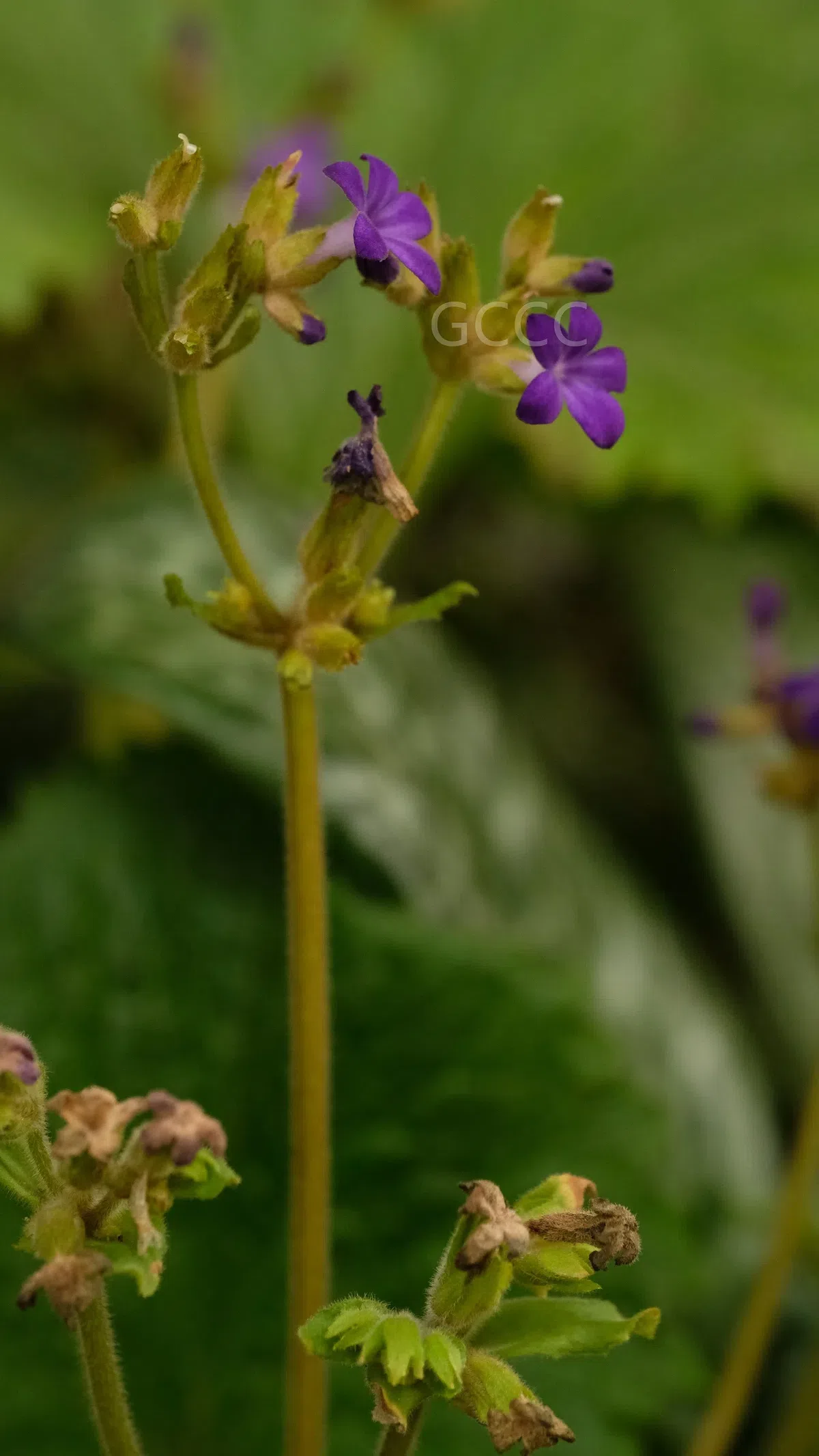
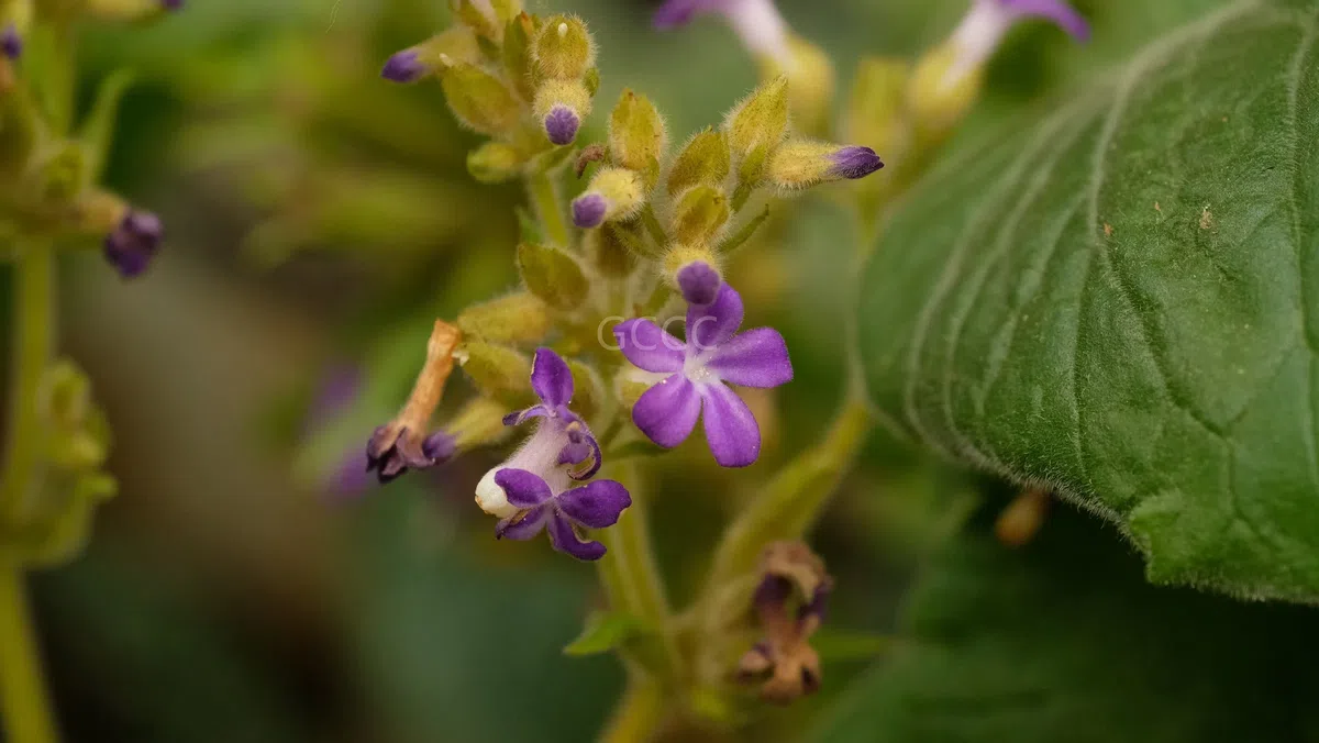
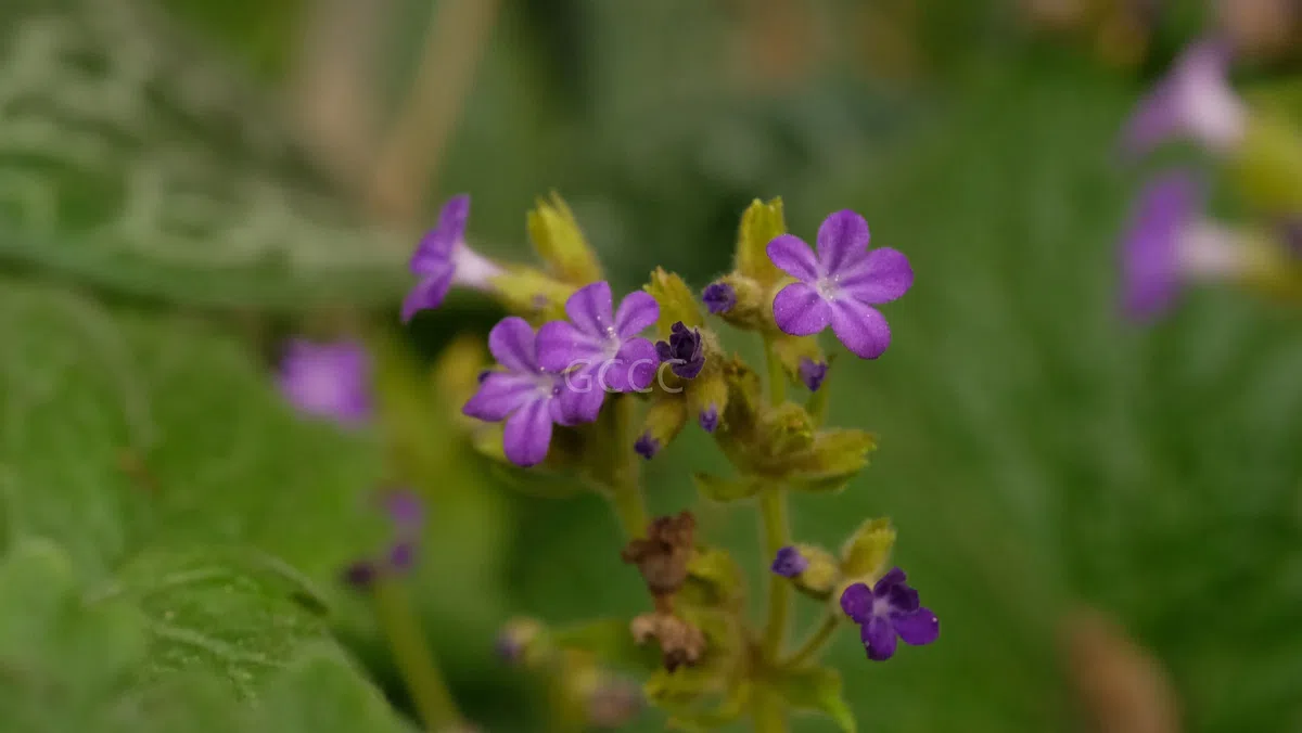
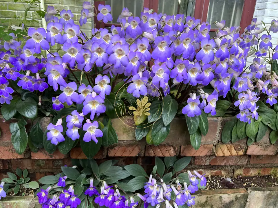
Figure 32-35.The flowering plants of Primulina tabacum in GCCC’s Nursery (Invasion?)