Kuan‑Ting Hsin, Chun‑Neng Wang
Expression shifts of floral symmetry genes correlate to flower actinomorphy in East Asia endemic Conandron ramondioides (Gesneriaceae)
Botanical Studies 2018 59:24
Background:
Bilateral symmetry flower (zygomorphy) is the ancestral state for Gesneriaceae species. Yet independent reversions to actinomorphy have been parallelly evolved in several lineages. Conandron ramondioides is a natural radially symmetrical species survived in dense shade mountainous habitats where specialist pollinators are scarce. Whether the mutations in floral symmetry genes such as CYC, RAD and DIV genes, or their expression pattern shifts contribute to the reversion to actinomorphy in C. ramondioides thus facilitating shifts to generalist pollinators remain to be investigated. To address this, we isolated putative orthologues of these genes and relate their expressions to developmental stages of flower actinomorphy.
Results:
Tissue specific RT‑PCR found no dorsal identity genes CrCYCs and CrRADs expression in petal and stamen whorls, while the ventral identity gene CrDIV was expressed in all petals. Thus, ventralized actinomorphy is evolved in C. ramondioides. However, CrCYCs still persists their expression in sepal whorl. This is congruent with previous findings that CYC expression in sepals is an ancestral state common to both actinomorphic and zygomorphic core Eudicot species.
Conclusions:
The loss of dorsal identity genes CrCYCs and CrRADs expression in petal and stamen whorl without mutating these genes specifies that a novel regulation change, possibly on cis‑elements of these genes, has evolved to switch zygomorphy to actinomorphy.
Link: https://doi.org/10.1186/s40529-018-0242-x
PDF Link: https://as-botanicalstudies.springeropen.com/track/pdf/10.1186/s40529-018-0242-x
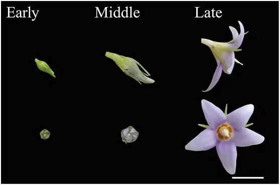
Fig. 1 The flower of C. ramondioides from early to late stage. Early, middle and late stage corresponding to Stage 10, 13, 15 in Additional file 1: Table S1. Actinomorphy was observed in late stage C. ramondioidesflower. Scale bar represented 1 cm
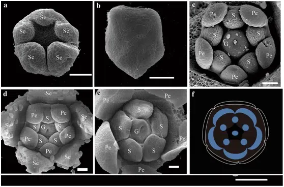
Fig. 2 The SEM photos of morphological development process of Conandronramondioidesflowers. Zygomorphy was observed at Stage 3 of sepal whorl. Definition of developmental stages of C. ramondioidesbased on (Harrison et al. 1999). a Stage 3, the sepals form as bulges at the points of the pentagon. b Stage 4, the sepals grow, while the floral meristem remains undifferentiated. c Stage 6, the corolla and androecium grow, and the gynoecium initiates. d Stage 7A, petal growth. e Stage 7B, androecium and gynoecium development. f Floral diagram of C. ramondioides. Se Sepals, Pe petals, S stamen, G gynoecium. Scale bar represents 50 μm
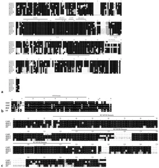
Fig. 3 Alignments of protein sequences of CrCYC,CrRADand CrDIVgenes with homologs from Antirrhinum majus(CYC , RAD and DIV) and Bournealeiophylla(BlCYC1, BlCYC2, BlRAD, BlDIV1 and BlDIV2).
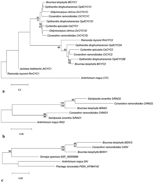
Fig. 4 Neighbour-joining trees of CYC -like, RAD-like and DIV-like genes.
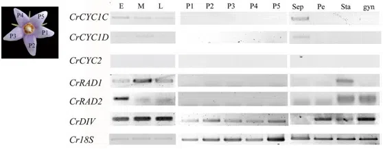
Fig. 5 Gene-specific reverse transcriptase polymerase chain reaction (RT-PCR) analysis of CrCYC,CrRADand CrDIVgenes from C. ramondioidesbuds and dissected flower tissues. E, M, L represent three flower development stage defined in this study. E stands for early flower development stage; M stands for middle flower development stage; L stands for anthesis stage. P1 to P5 represent dissected petal from flower bud at early (E) flower developmental stage. Sep, Pe, Sta and gyn denote pooled sepals, pooled petals, pooled stamens and gynoecium dissected from early flower developmental stage. CrCYC1C, CrCYC1D and CrCYC2 indicate expression of CrCYC1C, CrCYC1D and CrCYC2. CrRAD1 and CrRAD2 indicate expression of CrRAD1 and CrRAD2. CrDIVindicates expression of CrDIV. 18S is included as a positive control. CrCYC1C, CrCYC1D, CrRAD1, CrRAD2 and CrDIVare detected through flower development stages, only CrCYC2 is restricted through flower development stages. CrDIVis detected in all petals, whereas CrCYC1C, CrCYC1D, CrCYC2, CrRAD1 and CrRAD2 are restricted in petals. CrCYC1C and CrCYC2 were detected in pooled sepal tissue
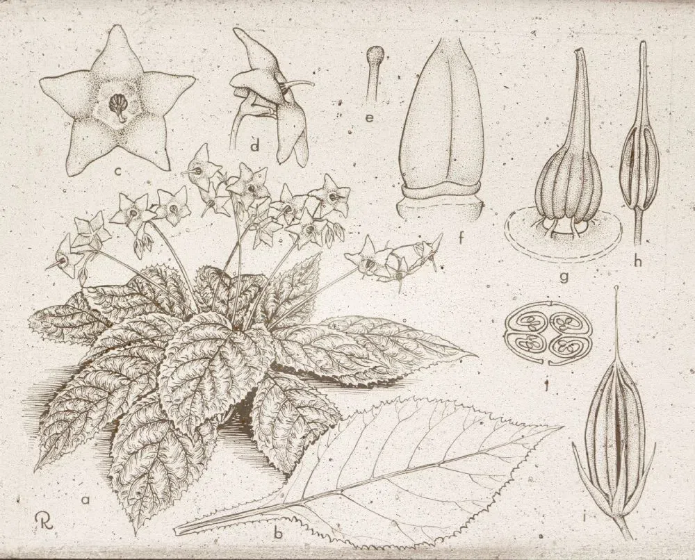
Fig.6 The drawing of Conandron ramondioidesSieb. &Zucc.
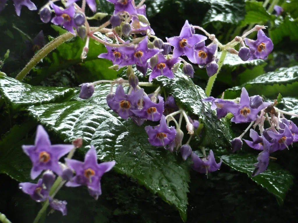
Fig 7
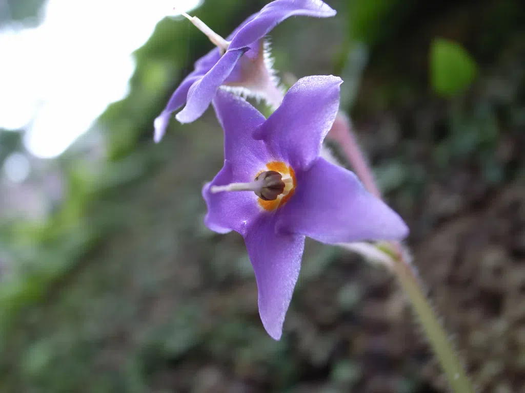
Fig 8
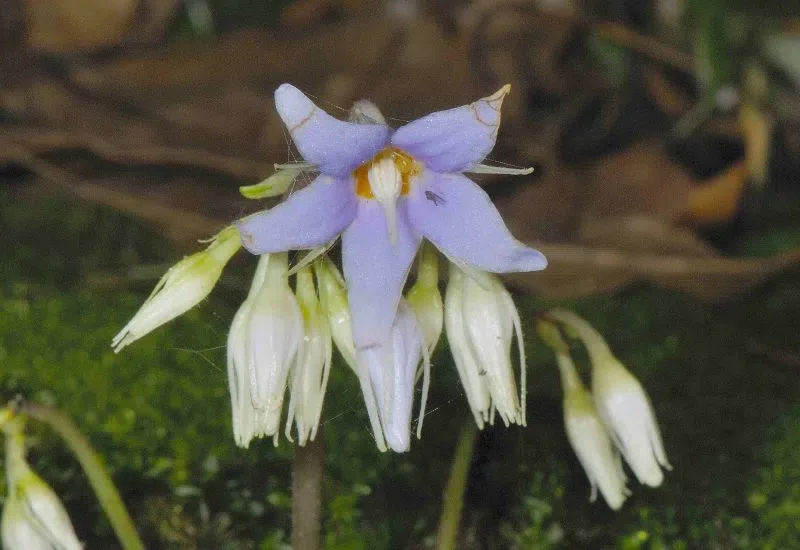
Fig 9
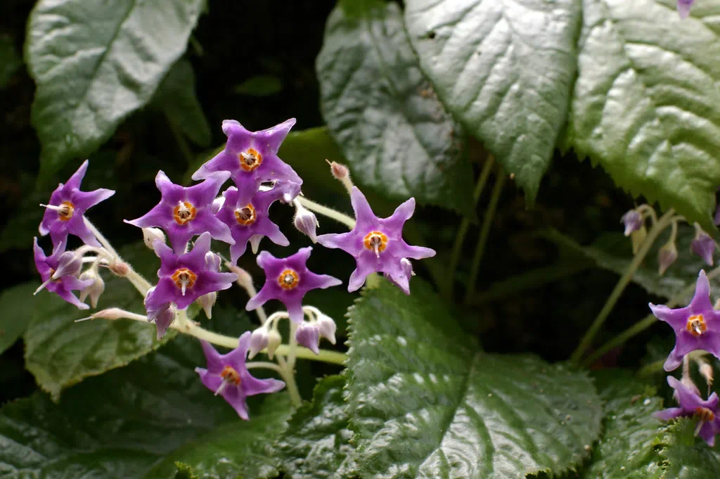
Fig 10
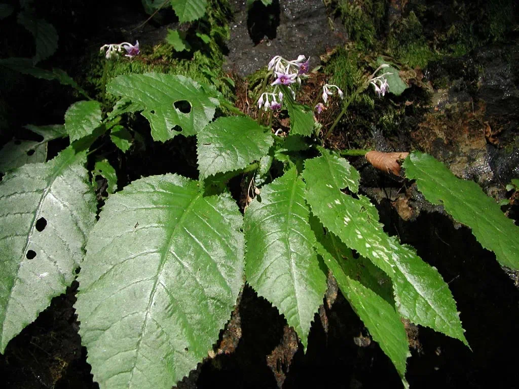
Fig 11
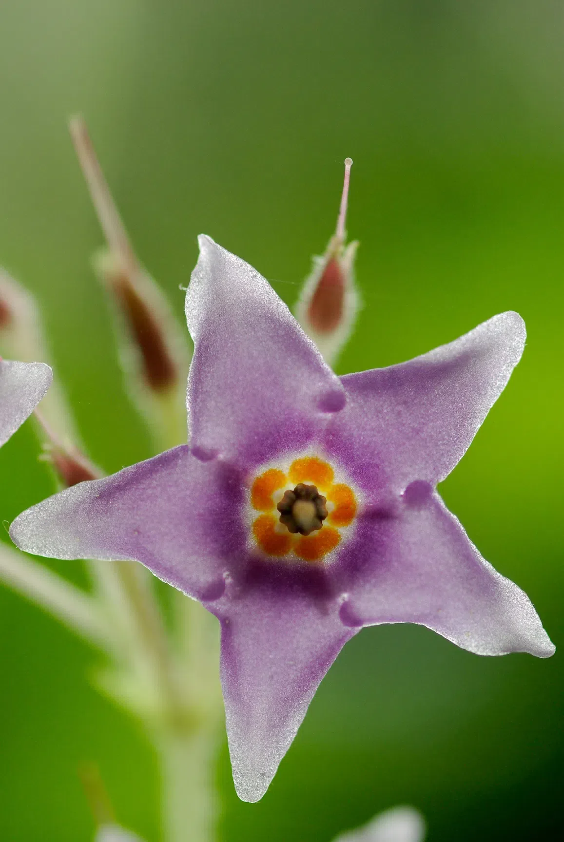
Fig 12
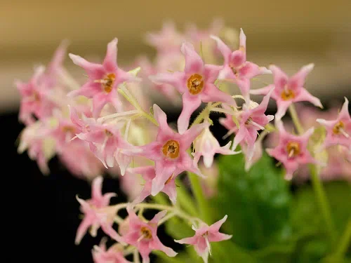
Fig 13
Fig. 7-13 Photos of Conand ronramondioides Sieb. &Zucc. (Cited from Flickr& Internet)


