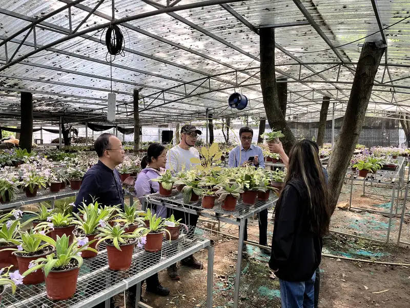

No.1 XIN HONG, Jeremy Keene, ZHI-JING QIU& FANG WEN*
Primulina anisocymosa (Gesneriaceae),
a new species with a unique inflorescence structure from
PeerJ 7:e6157 https://doi.org/10.7717/peerj.6157
ABSTRACT
A new Primulina
species from
Original article link:https://peerj.com/articles/6157
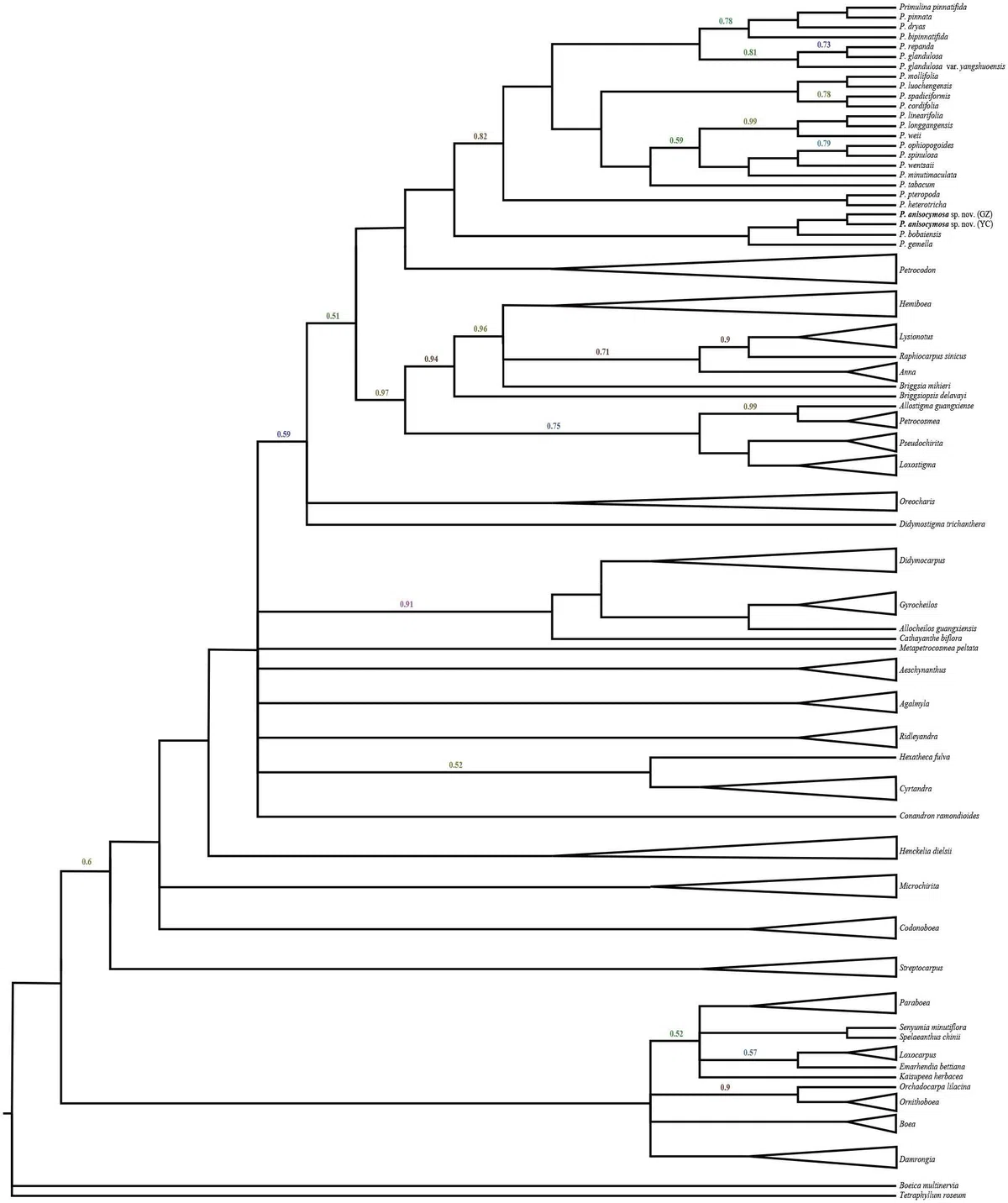
Figure 1 Molecular result showing two populations of this new species was strongly supported in a clade, and fell deeply nested in amonophyletic Primulina. Bayesian inference tree focusing on Primulina inferred fromcombined trnL-FCITS data, Bayesian posterior probabilities values less than 1.00 are labeledalong branches, all other nodes had PP values of 1.00. (GZ: typelocality, Gaozhou city; YC: Yangchun city).
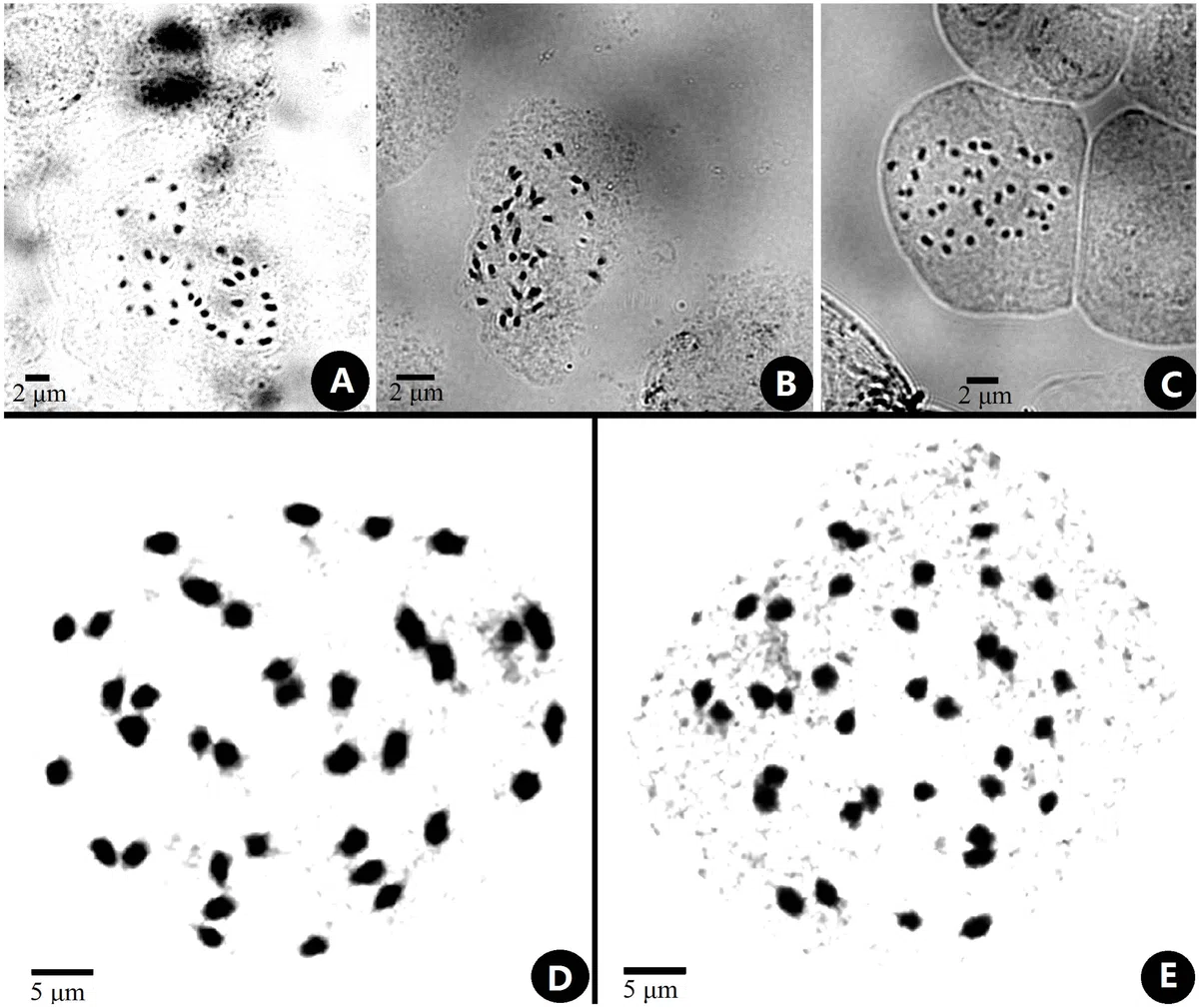
Figure 2 Somatic chromosomes at metaphase of Primulina anisocymosa. Somaticchromosomes at metaphase of Primulina anisocymosa (2n D 36, fromthe leaf cuttings). (A–C). Microphotograph from different cells (A, B:Gangzhou, E: Yangchun). D, E: Photomicrographs of mitotic metaphase chromosomes(D: Gaozhou, E: Yangchun). Photos by Ms. Sun-Lan Yin.
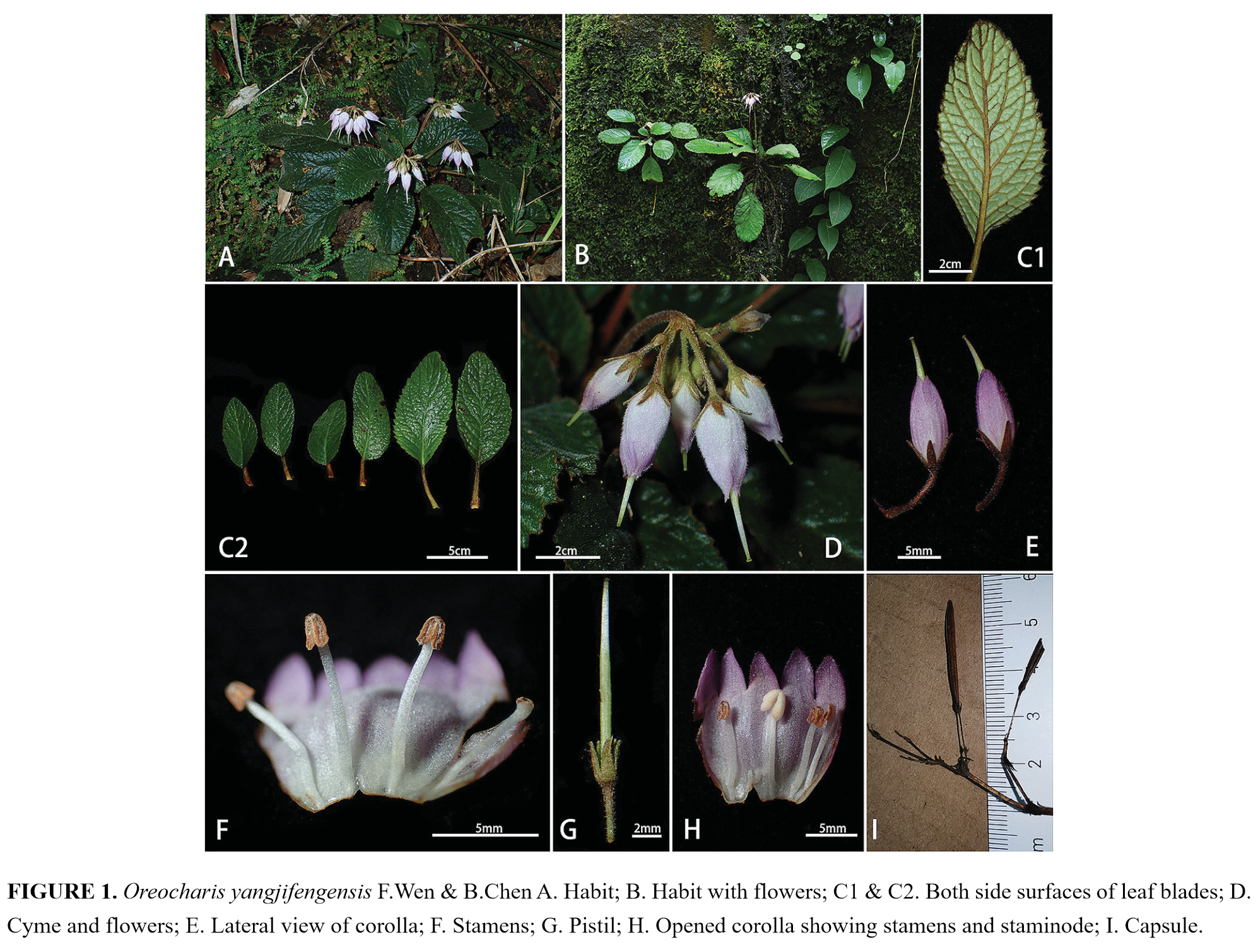
Figure 3 Line-drawing of Primulina anisocymosa. Primulina anisocymosa F. Wen, Xin Hong & Z.J. Qiu. (A) Plantin flower. (B) Corolla opened showing stamens and staminodes. (C) Calyx andpistil with ovary, style and stigma. (D) Seed. —Drawn by Ms. Xiao-Ming Xu andMs. Wen Ma.
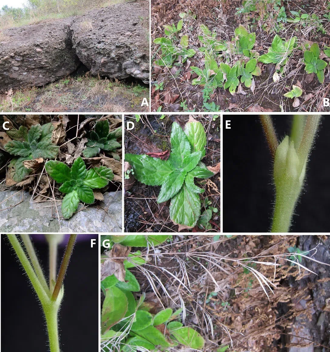
Figure 4 Photos of Primulina anisocymosa two populations in natural habitat. Primulina anisocymosa F. Wen, Xin Hong & Z.J. Qiu) (A, B) Habitat (A, Gaozhou; B,Yangchun) (C, D) Vegetative part of plants (C, Yangchun; D, Gaozhou) (E)Bracteoles, showing not-paired, aligned on one side at the base of pedicel. (F)Cymule, reduced to the point attachment and forming swollen nodules at thebase. (G) Zigzag monochasial infructescence. Photos by Fang Wen
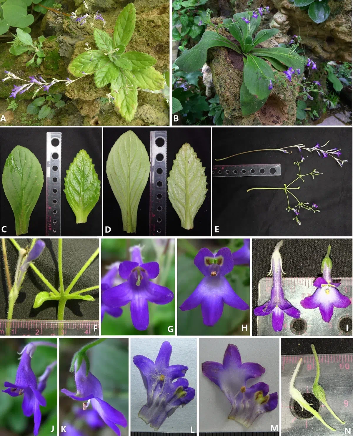
Figure 5 Comparison of Primulina anisocymosa and P. bobaiensis. Comparison of Primulina anisocymosa and P. bobaiensis,(A) Habit of P. anisocymosa when in flower. (B) A. Habit of P.bobaiensis when in flower. (C) Adaxial leaf blades (left: P. bobaiensis,right: P. anisocymosa). (D) Abaxial leaf blades (left: P. bobaiensis,right: P. anisocymosa). (E) Cymes (upper: P. anisocymosa, lower: P.bobaiensis). (F) Bracts (left: P. anisocymosa, right: P.bobaiensis). (G) Frontal view of corolla of P. anisocymosa. (H)Frontal view of corolla of P. bobaiensis. (I) Top view of corolla (left:P. anisocymosa, right: P. bobaiensis). (J) Lateral view ofcorolla of P. anisocymosa. (K) Lateral view of corolla of P.bobaiensis. (L) Opened corolla of P. anisocymosa. (M) Opened corollaof P. bobaiensis. (N) Pistils without corolla (left: P. anisocymosa,right: P. bobaiensis). Photoed by Xin Hong and Fang Wen.